| Standard Objectives |
4x/10x/20x/60x |
| (automated motorized two-position turret) |
Customization possible |
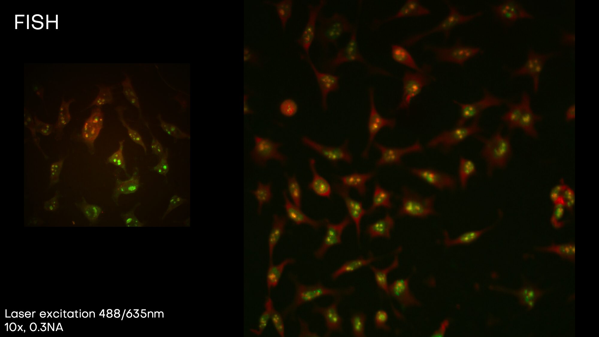
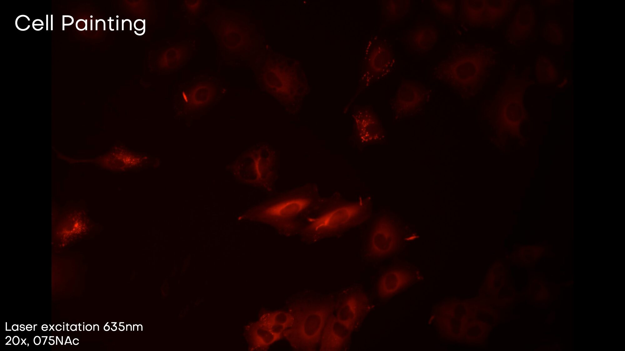
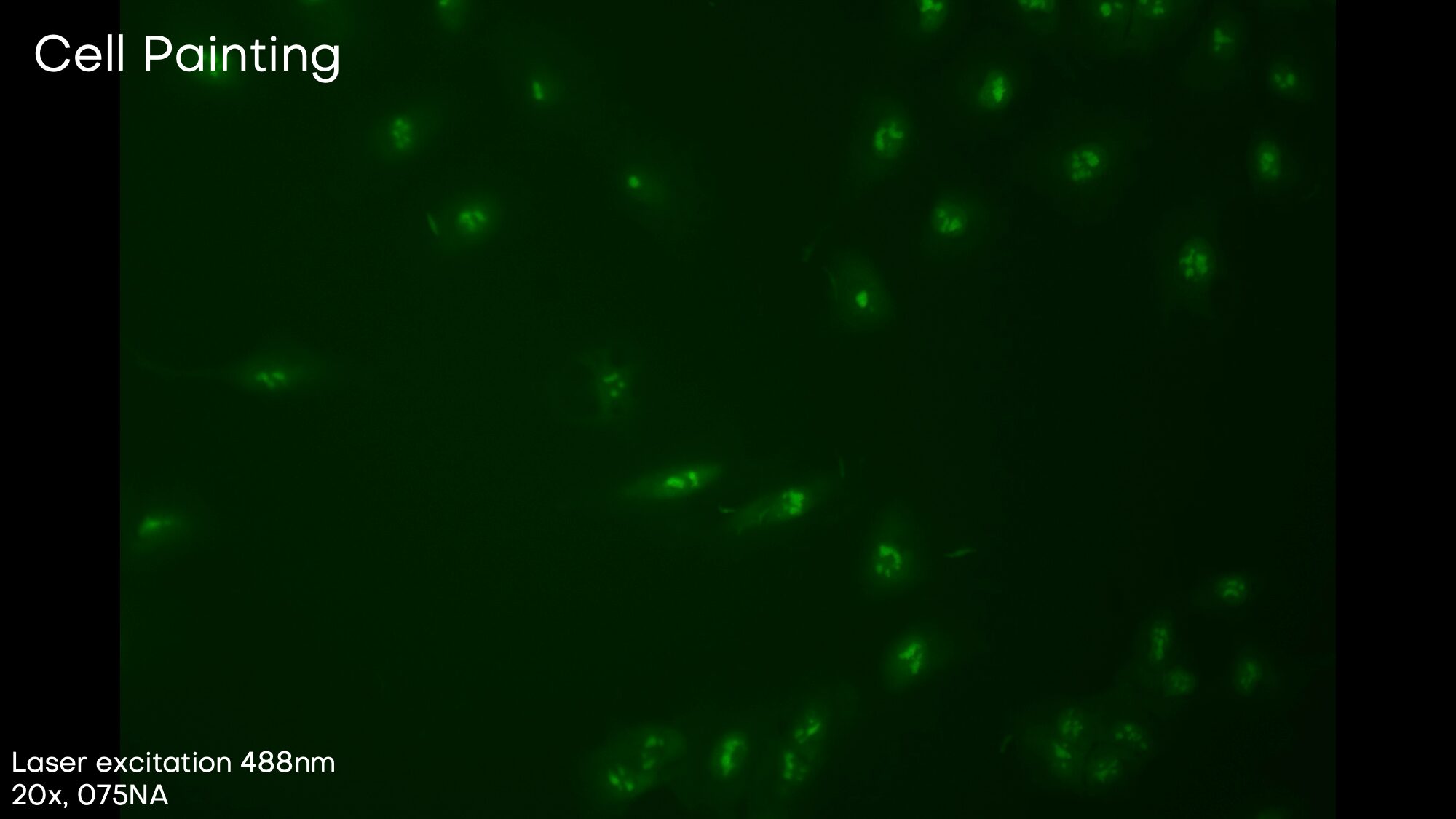
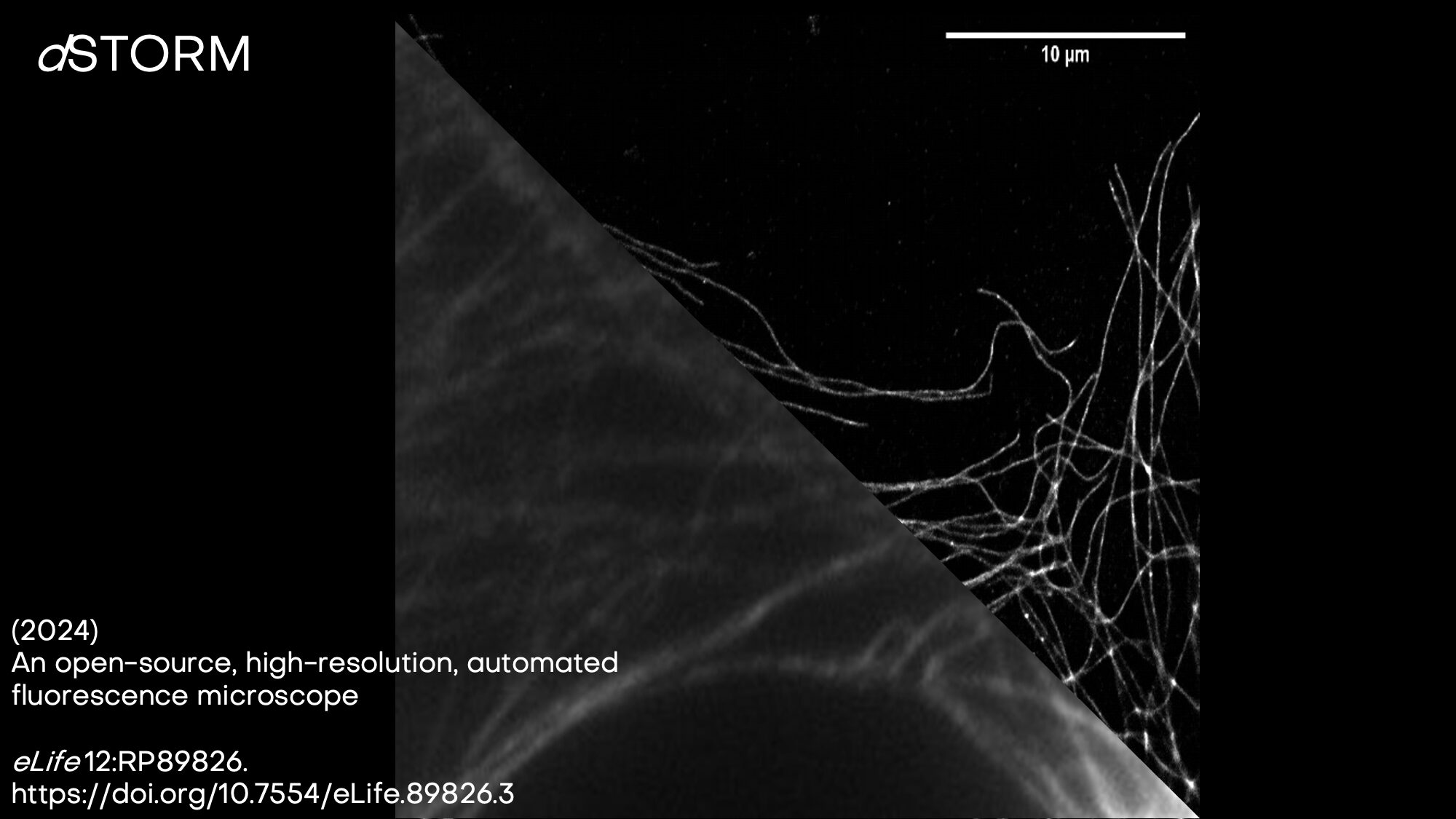
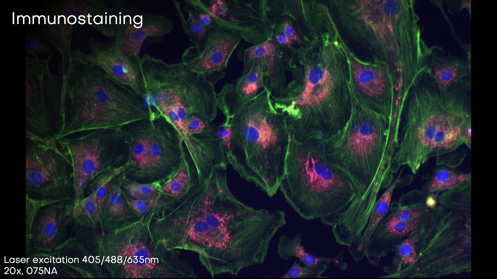
| Excitation Wavelengths |
405 nm (e.g. DAPI) |
| (single-laser, dual laser or quad laser configuration) |
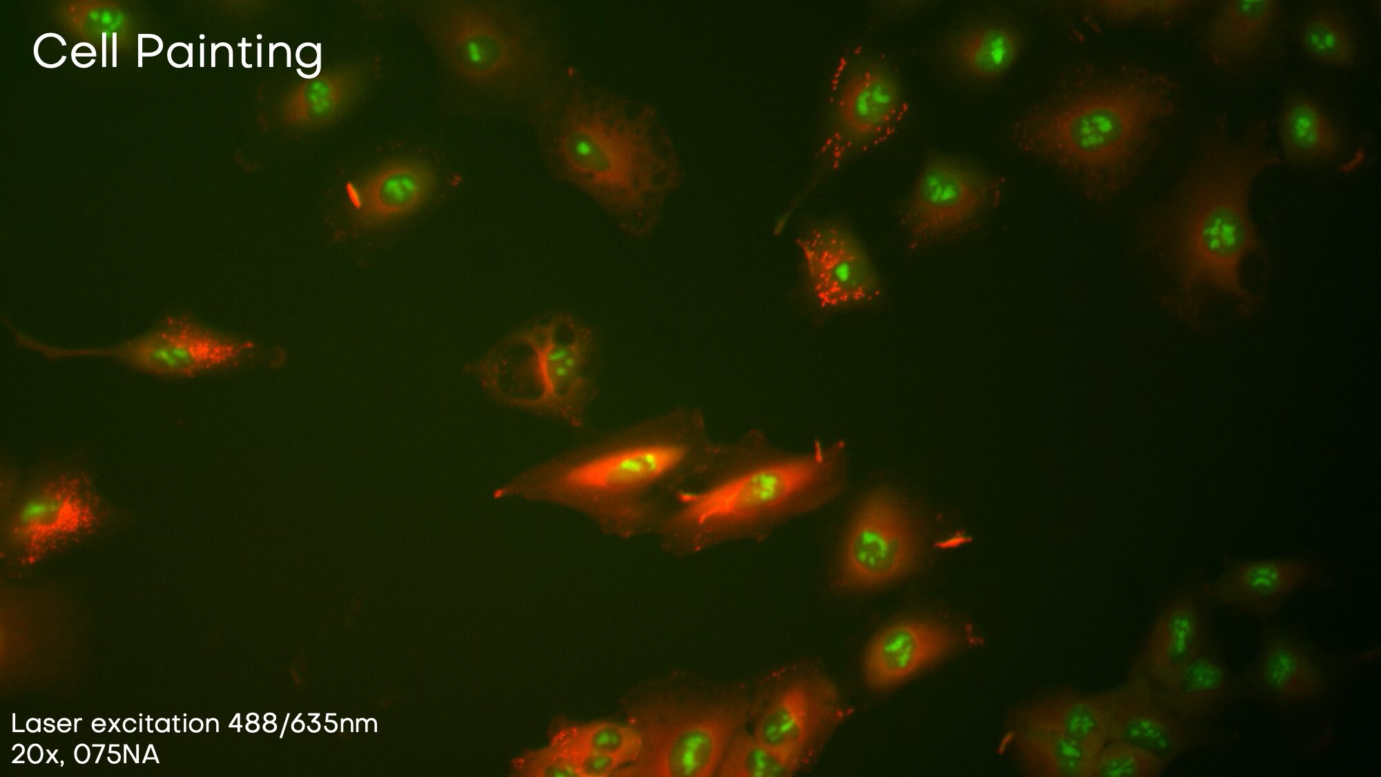
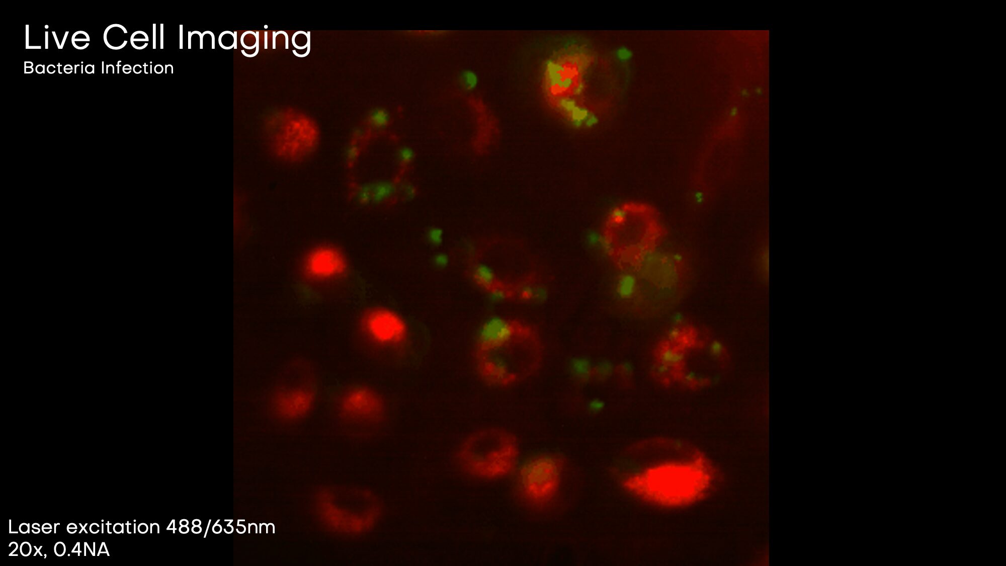
488 nm (e.g. GFP) |
| Emission Wavelengths |
520 nm, (e.g. mCherry) |
|
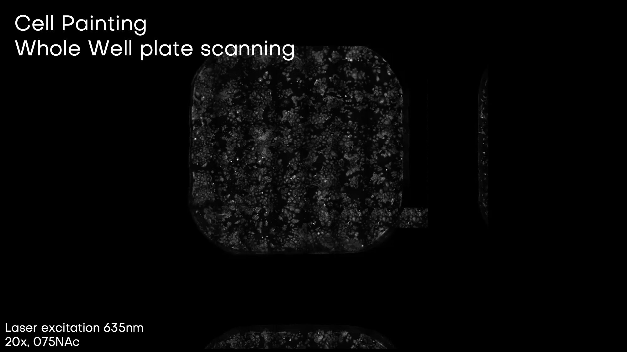
635 nm (e.g. AlexaFluor®647) |
|
Custom Cube Setup (typically DAPI 488 nm, GFP 520 nm, mCherry) 635 nm, AlexaFluor® 647) |
| Imaging Modes |
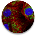
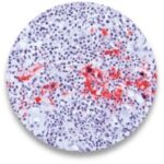
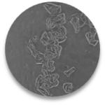
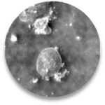
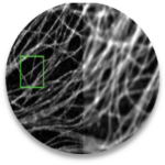
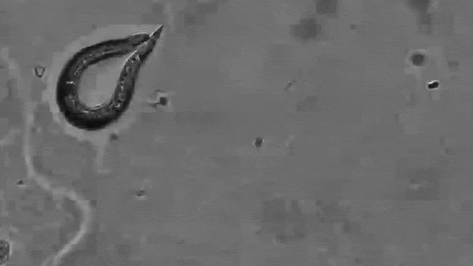
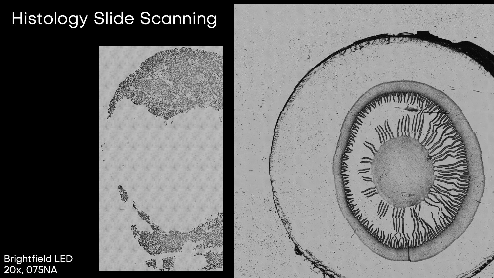
Brightfield, Colored Brightfield, Phase Contrast, Fluorescence |
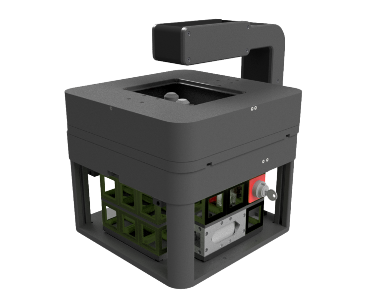
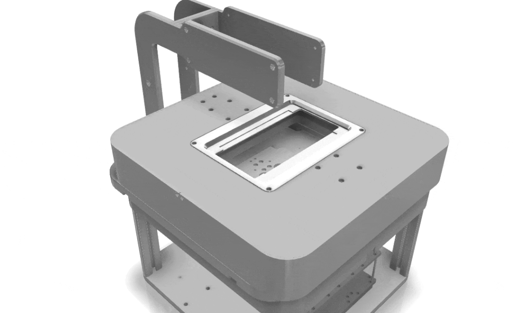
| Operating Range (XYZ) |
130 x 90 x 10 mm |
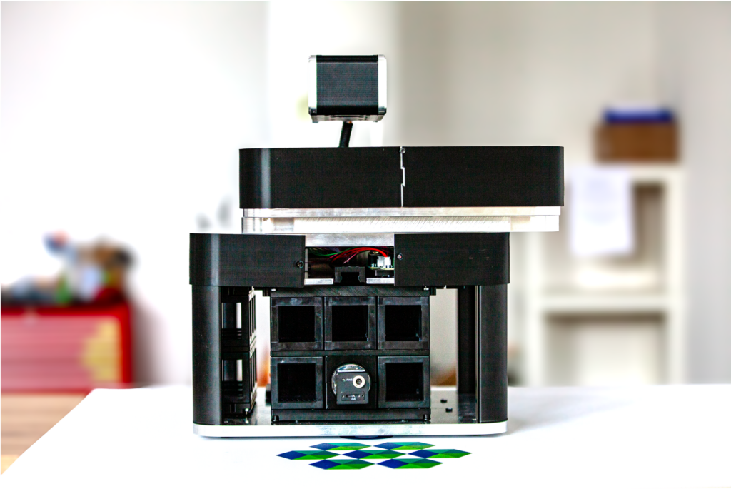
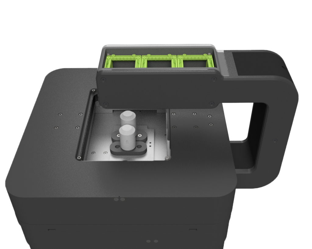
| Labware Supported |
Microplates (6x-384x), cell culture flasks (T25, T75), Petri- and cell culture dishes, microscope slides (4fold holder), count- and flow chambers, adapter for customized formats |
| Camera Compatibility |
Single/Dual-camera in cube assembly (e.g. RGB for Histology, Monochrome for Fluorescence), overview camera top |
| Optical Interface |
UC2 Cube system in 3x3x2 grid, 3x1x2 from the sides or 3x1 from above |
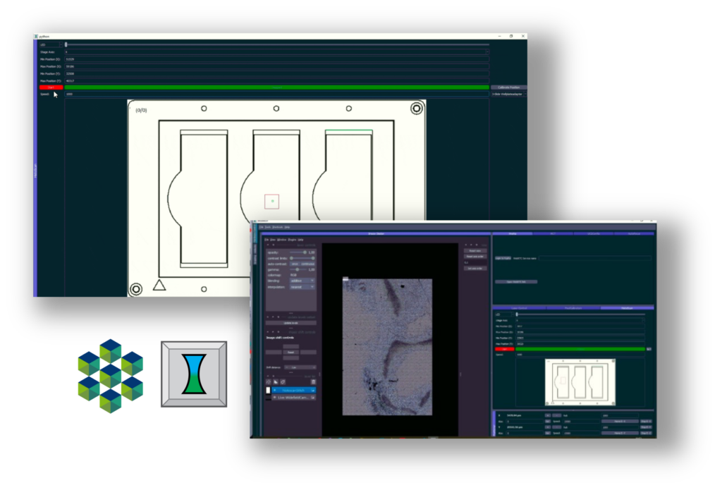
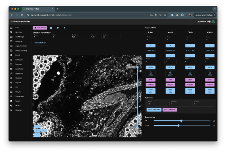
| Control Software |
ImSwitch running in Docker self-hosted or on internal Raspberry Pi; REST-API; USB Serial and Camera driver (HIK Robotics), PS Controller |
| Data Format |
OME-ZARR, OME-TIF, JPEG, MP4, PNG, customized |
| Software Interface (SDK & API) |
Comprehensive, for integration and automation |
| Dimensions & Weight |
300x300x300 mm3, 11, <16 Kg (without Top Illumination part) |
| Electric Input |
Power Supply: 110/230V, 50/60Hz |
|
12V, 5A (optional: battery-driven) |
| Environmental Conditions |
10-40°C, <95% humidity |

