
How the University of Helsinki built an open brightfield scanner for €15.000
Digital pathology increasingly depends on whole-slide imaging (WSI). But the scanners that produce it are priced for clinical throughput, not for research. For a lab running exploratory or early-stage biomarker work rather than routine diagnostic volume, that math never closes. Slide digitization either stays out of reach, or it means queuing for a slot at a shared core facility.
Cost is not only the purchase price though. Published figures put annual service contracts for commercial scanners at 7 to 20 percent of the instrument cost. Therefore a closed scanner is not a one-time purchase. Instead, at the upper end of that range, service alone matches the initial investment within five years.
The second cost is harder to put a number on. Even though proprietary formats can usually be read by open tools such as QuPath, it only works because projects like OpenSlide and Bio-Formats reverse-engineer them. That support lags behind new instrument generations, degrades once you move past plain brightfield RGB, and comes with no guarantee from anyone. A lab that builds a five-year image archive on a closed format is making a bet on volunteers keeping up with a vendor who has no interest in helping them.
That was the situation the University of Helsinki brought to us.
What we built
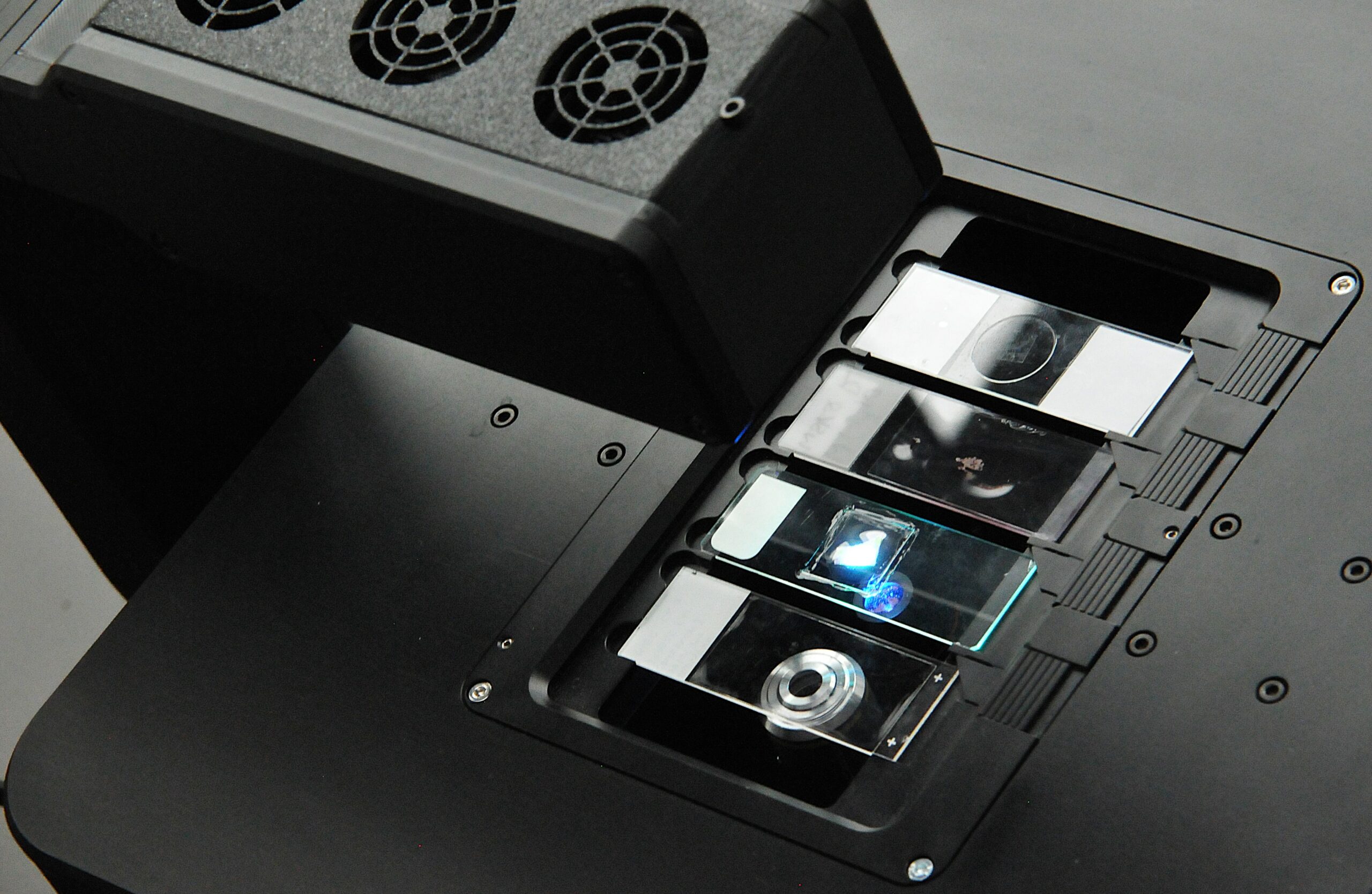
We supplied a Brightfield-FRAME configured for whole-slide scanning. Every FRAME is built on openUC2’s cube-based optical architecture: illumination, filter and detection paths are discrete modules – some 3D-printed, some off-the-shelf – that snap together on a standard grid, while our ImSwitch software makes sure they also talk to each other on the digital side.
For Helsinki’s use case, the configuration came down to four pieces:
- A motorized XYZ stage and application-specific optics tile across the full slide in transmitted brightfield light.
- Focus-mapping compensates for uneven mounting and slide thickness, holding focus across the entire tissue area.
- ImSwitch drives acquisition; ashlarUC2 stitches the tiles into a single whole-slide image with sub-pixel accuracy.
- Output is written in open, standard formats such as OME-TIFF, allowing further analysis in open tools.
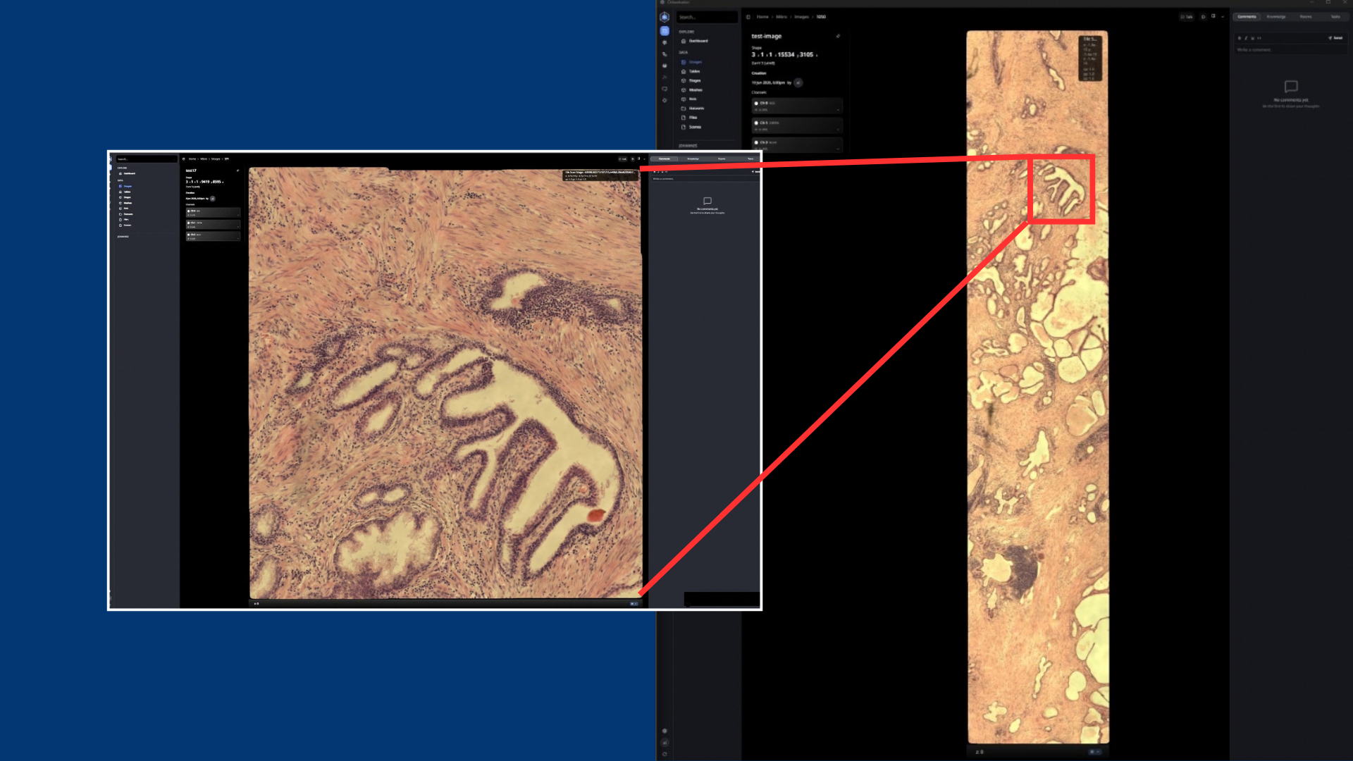
Einzelaufname (links) und zugehörige whole-slide-Aufnahme (rechts)
What it changed
The system has been running in Helsinki since June 2026.
“We used the FRAME in our lab for various automation workflows. In contrast to other systems used for automation, we can also use it quickly and without fuss manually, i.e. to scan individual slides to check their quality before we continue working with them.
This saves precious time in the overall workflow since we don’t need to configure our FRAME but can instead instantly start to scan the slides!”

Franziska Niemeyer
But €15.000 is not the number that matters
What actually matters is the cost of the next experiment. Helsinki now owns a base platform, not a fixed-function instrument. The same mechanical FRAME and the same ImSwitch stack that scan brightfield IHC today can be reconfigured (in-house, with our help or with material from someone else entirely) for a different use case tomorrow. This brings down the cost for the next experiment with no vendor lock-in.
That is also how this story continues. The next case study picks up where this one ends: the same FRAME, this time inside a fully automated, closed-loop multiplex staining pipeline